Stand-Up MRI of Ft. Lauderdale
4616 North Federal Highway
Ft. Lauderdale, FL, 33308
Tax ID: 65-0637743 NPI: 1518059781
Check your email once you have been scheduled or scanned for a username and password
MRI Case Studies
There is considerable clinical evidence that Weight-Bearing UPRIGHT® MRI provides medical benefits that are not duplicated by any other MRI
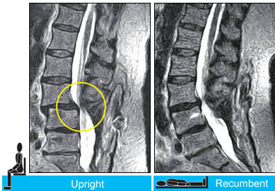
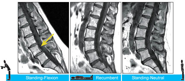
Case #1: Postoperative Hypermobile Instability
You need the upright scan to see the spinal instability in this patient with recurrent low back pain following an L4-S1 fusion.
(Images courtesy of M. Rose, M.D., Rose Radiology Centers)
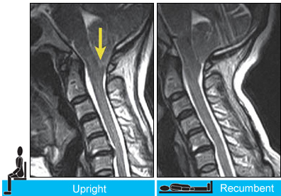
Case #2: Chiari Malformation Visualization When Upright
Upright imaging revealed increased downward herniation of the cerebellar tonsils (arrow). Subsequent neurosurgery (a posterior fossa decompression) eliminated the patient's sudden drop attacks.
(Images courtesy of J.P. Elsig, M.D., Zurich, Switzerland)
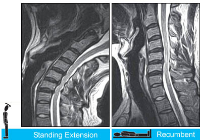
Case #3:Upright Dynamic MRI Reveals Hidden Disc Herniation
You need the upright scan to see the position-dependent focal posterior disc herniation at the C4/5 level (arrow). Note the asscociated spinal cord compression on the standing-extension scans in both the sagittal (top left) and axial planes(bottom left).
(Images courtesy of Melville MRI, P.C.)
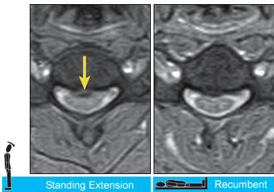
Case #4: Evaluation of Spinal Instability
You need the upright scan to see the position-dependent focal posterior disc herniation at the C4/5 level (arrow). Note the asscociated spinal cord compression on the standing-extension scans in both the sagittal (top left) and axial planes(bottom left).
(Images courtesy of Melville MRI, P.C.)